Clinical UpdateSeptember 1, 202603 / 03
Hunan Huachuang Medical Technology Co., Ltd.
Latest clinical updates
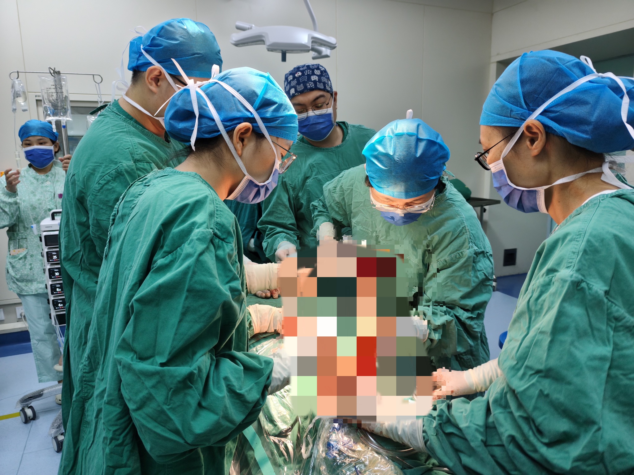
On September 1, 2026, The First Affiliated Hospital of University of South China used HC-IORT to successfully complete Hunan’s first intraoperative radiotherapy procedure for oropharyngeal cancer. Treatment field diameter: 30 mm; prescribed dose: 10 Gy; energy: 6 MeV; tissue bolus: 10 mm; beam-on time: 3.03 minutes.

Rednet News, September 4 (Correspondents: Wu Fang and Yang Dong) — Recently, The First Affiliated Hospital of University of South China achieved an important technical breakthrough in oral cancer treatment by successfully applying Hunan’s first domestically developed mobile electron beam intraoperative radiotherapy (IORT) system in clinical care for oral cancer. This provides patients with a new, integrated treatment option combining surgery and precision radiotherapy. The hospital had already established the use of this technique in breast cancer, colorectal cancer and other cancers, accumulating extensive experience. Its successful extension to oral cancer further broadens the hospital’s IORT applications and advances its capabilities in precision cancer care. The domestically developed mobile electron beam IORT system offers high precision, adaptability and safety. It supports three electron beam energies — 6 MeV, 9 MeV and 12 MeV — with a treatment depth range of 0–40 mm and a typical dose rate of 300–400 cGy/min. Available field sizes include 1 cm and 3–10 cm planar fields, as well as 4 mm and 10–50 mm spherical fields. Combined with automated soft docking and positioning, the system can deliver a single, precisely targeted high dose to the tumor bed immediately after tumor resection, effectively eliminating residual cancer cells while maximizing protection of surrounding normal tissues and organs and avoiding the drawbacks of conventional radiotherapy. The patient treated with this new approach had high-risk oral cancer. Although the primary lesion in the oral cavity was small, there were multiple ipsilateral cervical lymph node metastases, with the largest node measuring 4 cm and lying immediately adjacent to the internal carotid artery and internal jugular vein. The risk of postoperative recurrence was high, and the prognosis with conventional surgery was limited. To minimize recurrence risk and optimize treatment outcomes, the surgical team led by Deputy Director Lü Jie of the Department of Stomatology promptly convened a multidisciplinary team (MDT) consultation with the radiotherapy team led by Director Manbo Cai of the Department of Oncology, Associate Chief Physician Chen Lixun of the Department of Medical Imaging, Associate Chief Physician Yang Shuai of the Department of Pathology and other specialists. Following careful assessment, the team developed an individualized combined treatment plan of wide excision of the lesions followed by precision IORT. During surgery, the medical team first completely removed the primary oral tumor and the metastatic lesions in the neck. To address the risk of high-dose radiation damaging nearby nerves, the surgical and radiotherapy teams worked closely together to fully mobilize the nerves adjacent to blood vessels within the irradiation field and carefully shield them with lead tubing and lead sheets. This enabled targeted high-dose irradiation of the tumor bed under direct visualization to precisely eliminate potential residual disease while safeguarding normal tissues. From conventional radiotherapy to targeted tumor clearance with IORT, and from breast and colorectal cancers to oral cancer, the hospital’s team has consistently used technological innovation to advance cancer care. The successful application of IORT to oral cancer demonstrates the hospital’s core capabilities and innovation in precision oncology as a regional medical center, helping fulfill its commitment to enabling patients with serious illnesses to receive care within Hunan. The hospital will continue to advance cutting-edge medical technologies and pursue clinical excellence, translating high-quality, precise care into health benefits for people across the province.
Hunan Daily · New Hunan, September 15 (Correspondent: Long Hong) — Recently, the gastrointestinal surgery team led by Ouyang Jun and the oncology team led by Manbo Cai at The First Affiliated Hospital of University of South China jointly performed resection of a recurrent tumor combined with intraoperative radiotherapy (IORT) for a patient whose right retroperitoneal liposarcoma had recurred after multimodal treatment. This was the fifth liposarcoma IORT case completed through collaboration between the two teams, adding to their clinical experience in the comprehensive treatment of complex retroperitoneal tumors. The patient had previously undergone surgery for liposarcoma and systemic chemotherapy and sought care at the hospital after the tumor recurred. Because the recurrent tumor lay deep, was adjacent to major blood vessels, involved the psoas major muscle and had complex anatomical relationships, a repeat operation would be challenging. Ouyang Jun’s and Manbo Cai’s teams jointly assessed the case and devised a plan to complete tumor resection and precision IORT during the same operation. During surgery, Ouyang Jun’s team carefully separated the tumor from surrounding tissues and completely resected the recurrent tumor and involved areas while protecting major vessels, nerves and adjacent organs. Immediately after resection, Manbo Cai’s team delivered IORT to the tumor bed at high risk, directing radiation to the local area most in need of additional control and seamlessly combining surgery with precision radiotherapy. For abdominal tumors, a major advantage of IORT over conventional external beam radiotherapy is that normal tissues and organs can be displaced outside the treatment field under direct surgical visualization. This helps spare them from irradiation while delivering an effective tumoricidal dose to the target and concentrating treatment on the area at high risk, thereby strengthening local treatment while minimizing exposure of surrounding normal tissues as far as possible. The two teams have now jointly completed five liposarcoma IORT cases. Each case underwent rigorous preoperative assessment, multidisciplinary discussion and radiotherapy equipment quality assurance (QA) checks and was completed using a standardized workflow. The cases provide preliminary indications of good safety and feasibility and offer a new combined treatment option for patients with complex or recurrent retroperitoneal tumors. Experts note that retroperitoneal liposarcomas are often deep and large, lie close to major blood vessels, nerves and organs, and carry a relatively high risk of local recurrence. For patients with recurrent disease in particular, repeat surgery alone may still leave local control inadequate. Combining surgical resection with precision IORT allows the high-risk tumor bed to be irradiated directly at close range after resection. Together with preoperative or postoperative radiotherapy, the approach aims to improve local tumor control and overall survival. From their initial case to the fifth liposarcoma IORT procedure, Ouyang Jun’s and Manbo Cai’s teams at the hospital have continued refining the coordination between surgery and radiotherapy. The patient’s decision to seek care there and successful completion of treatment also reflect the hospital’s growing capabilities in surgery, radiotherapy and multidisciplinary management of complex retroperitoneal tumors.
Scroll down to view earlier news
Rednet News, August 28 (Correspondent: Long Hong) — Giant retroperitoneal liposarcoma is widely regarded as one of the most difficult challenges in abdominal surgery. These tumors lie deep in the abdomen and may encase or compress major blood vessels and vital organs, with any misstep risking irreversible organ damage. Recently, the gastrointestinal surgery team led by Ouyang Jun at The First Affiliated Hospital of University of South China completely removed a massive right retroperitoneal liposarcoma occupying two-thirds of the abdominal cavity. Under exceptionally demanding conditions, the team preserved the patient’s only functioning right kidney and worked with Manbo Cai’s radiotherapy team from the Department of Oncology to deliver intraoperative radiotherapy to the tumor bed, strengthening protection against recurrence. The tumor was enormous and had displaced the abdominal organs forcefully to the left. The situation was even more critical because the patient’s left kidney had previously become completely atrophic due to stones, leaving the right kidney as the only functioning kidney. The giant tumor was densely adherent to the right renal capsule, with poorly defined boundaries of invasion. The surgical team faced a severe dilemma: removing the right kidney together with the tumor to maximize clearance would immediately leave the patient dependent on lifelong dialysis, while forceful dissection risked injuring the renal vessels or kidney tissue. Major bleeding or impaired renal blood supply could likewise result in the loss of the patient’s only functioning kidney. The conflict between preserving the kidney and achieving radical tumor removal left almost no margin for error. To overcome this challenge, gastrointestinal surgery convened a multidisciplinary team with oncology, urology, vascular surgery, anesthesiology, nephrology and the operating room before surgery. The team repeatedly reviewed CT images, evaluated the boundaries between the tumor and the renal vessels, inferior vena cava and bowel, and established two plans: strive for complete tumor separation while preserving the right kidney, while also preparing emergency responses for major bleeding or renal injury and finalizing the intraoperative radiotherapy plan. In the operating room, the tumor had displaced normal anatomy and made exposure difficult. The surgeons carefully separated the tumor little by little from the bowel and major retroperitoneal vessels. In the high-risk area where the tumor adhered to the right kidney, they meticulously distinguished the tumor pseudocapsule from renal tissue, avoided the renal blood vessels and combined blunt and sharp dissection. They sought to remove the tumor completely while preventing tears to the kidney tissue or injury to the renal pedicle. Through precise work, the team successfully removed the giant liposarcoma intact and preserved the patient’s only functioning right kidney. Immediately after the specimen was removed, Manbo Cai’s radiotherapy team delivered intraoperative electron radiotherapy to the tumor bed at high risk of recurrence and the region surrounding the right kidney where the tumor had been adherent. The open surgical field enabled precise targeting of areas at risk for residual tumor cells, reducing the risk of local recurrence and seamlessly integrating surgical resection with radiotherapy without a second operation or anesthetic. Retroperitoneal liposarcoma usually produces few specific symptoms at an early stage. By the time patients seek care for abdominal distension or enlargement, the tumor is often already massive, prone to invading nearby organs and associated with a high rate of postoperative local recurrence. The difficulty of this case lay not only in removing the giant tumor but also in balancing radical cancer treatment with preservation of the patient’s only functioning kidney, placing exceptional demands on anatomical expertise and intraoperative judgment. The successful operation demonstrates the hospital’s capability in managing complex retroperitoneal tumors. Through multidisciplinary collaboration, the team integrated surgery and IORT into a unified treatment approach, providing a stronger option for patients with complex retroperitoneal tumors in southern Hunan. The patient currently has stable vital signs and kidney function and is recovering well.
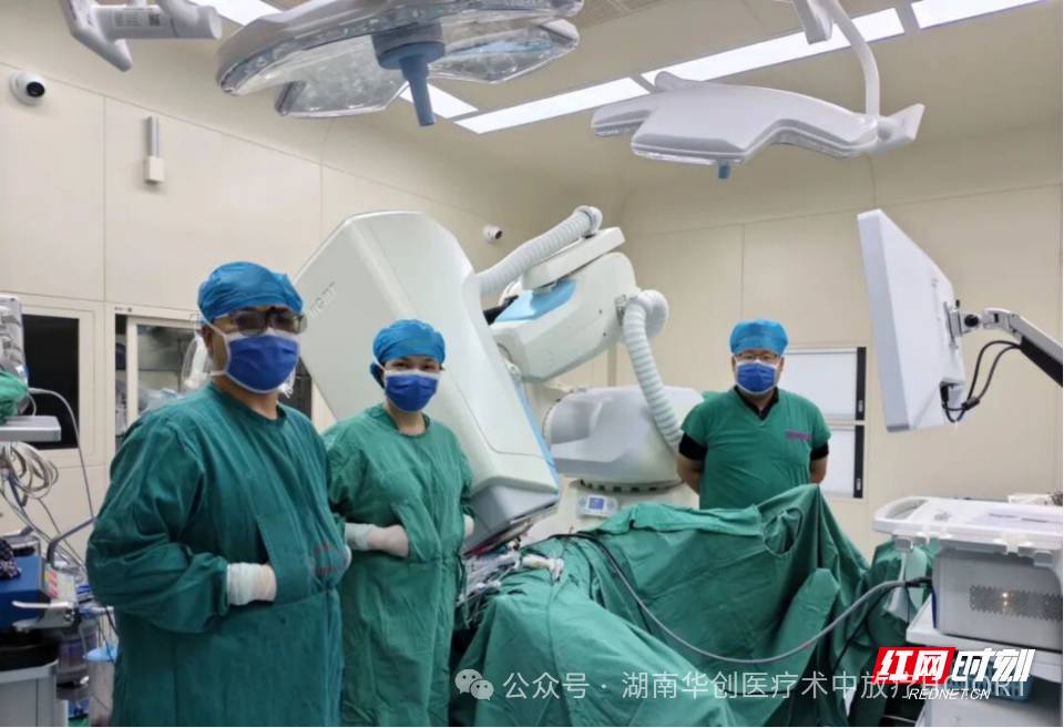
Rednet News, August 24 (Correspondent: Long Hong) — Recently, the gastrointestinal surgery team led by Ouyang Jun at The First Affiliated Hospital of University of South China, working with the radiotherapy team led by Manbo Cai from the Department of Oncology, successfully performed a 3D laparoscopic radical resection for rectal cancer (Dixon procedure). Using HC-IORT, the teams completed the hospital’s first rectal cancer intraoperative radiotherapy procedure with an electron beam spherical applicator, seamlessly integrating surgical resection with IORT. Rectal tumors are located within the narrow, deep pelvis. In some high-risk patients, subclinical disease that is difficult to detect visually may remain even after all visible tumor has been surgically removed, creating a risk of local recurrence. IORT can deliver a localized boost to areas at high risk of recurrence during surgery. Normal tissues can be displaced outside the irradiation field for protection, allowing potential residual disease to be targeted precisely. When combined with surgery and postoperative external beam radiotherapy, this approach may further improve surgical benefit and the likelihood of cure. During this procedure, Ouyang Jun’s team used 3D laparoscopy to complete precise radical resection of the rectal tumor. After tumor removal, Manbo Cai’s radiotherapy team identified the target area at high risk of recurrence under direct laparoscopic visualization. Because the pelvic surgical bed may be curved, recessed or otherwise irregular, a conventional planar applicator may not conform adequately or match the intended treatment area. The clinical team therefore selected a 50 mm electron beam spherical applicator. Its curved surface conformed closely to the complex deep-pelvic surgical bed, reducing geometric mismatch and helping deliver an adequate, uniform dose to the tumor bed. The prescribed dose was 15 Gy using a 9 MeV electron beam, with an actual beam-on time of only 4.91 minutes. Precise resection and targeted irradiation were completed during the same operation, providing a new technical approach to local treatment of high-risk rectal cancer in the deep pelvis. The successful procedure represents another advance in multidisciplinary rectal cancer care at the hospital and may help strengthen comprehensive treatment capabilities for gastrointestinal tumors in southern Hunan.
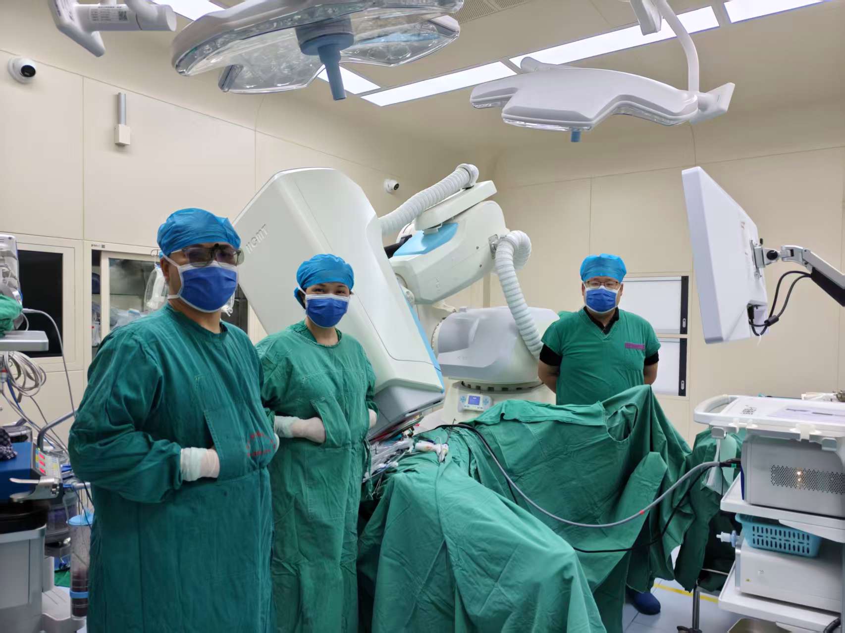
On August 18, 2026, The First Affiliated Hospital of University of South China used HC-IORT to successfully complete the world’s first rectal cancer IORT procedure with an electron beam spherical applicator. Spherical treatment field diameter: 50 mm; prescribed dose: 15 Gy; energy: 9 MeV; beam-on time: 4.91 minutes.
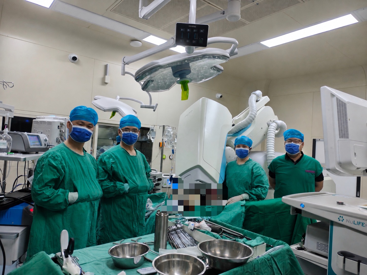
Following the world’s first laparoscopic IORT procedure for rectal cancer using a laparoscopic applicator on August 10, The First Affiliated Hospital of University of South China completed another minimally invasive laparoscopic rectal cancer IORT procedure on August 13, 2026. HC-IORT and a laparoscopic applicator were introduced through a trocar. Two laparoscopic treatment fields were delivered, and fluorescence guidance was used for the first time to assist IORT. Laparoscopic treatment field 1 — prescribed dose: 12 Gy; energy: 6 MeV; field diameter: 30 mm; field angle: 30°; beam-on time: 3.68 minutes. Laparoscopic treatment field 2 — prescribed dose: 10 Gy; energy: 6 MeV; field diameter: 30 mm; field angle: 30°; beam-on time: 2.98 minutes.
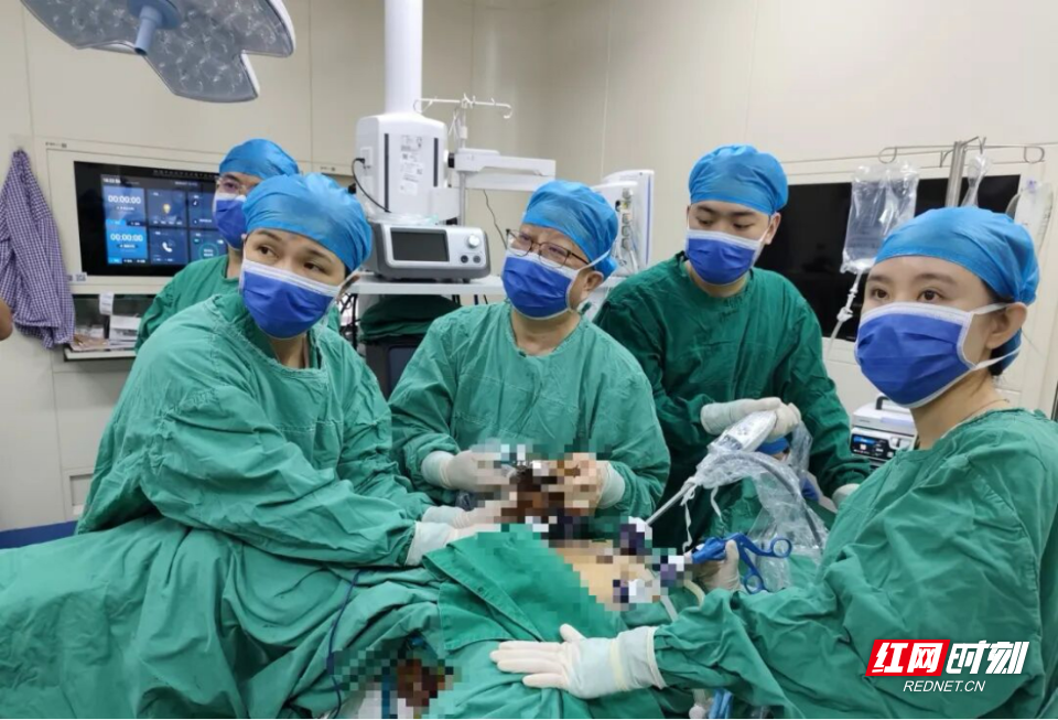
Rednet News, August 12 (Correspondent: Long Hong) — On August 10, the gastrointestinal surgery team led by Ouyang Jun and the radiotherapy team led by Manbo Cai at The First Affiliated Hospital of University of South China worked closely together to complete the first laparoscopic intraoperative radiotherapy (IORT) procedure for rectal cancer using HC-IORT, the first system of its kind in China and independently developed in Hunan. The operation integrated transanal total mesorectal excision (TaTME), lateral lymph node dissection and IORT. After radical tumor resection, ultra-low sphincter preservation and lateral lymph node dissection, a single precise high dose of radiation was delivered to the area at risk for subclinical disease. Surgery and radiotherapy were therefore completed in the same operating session, providing a more precise and individualized multidisciplinary treatment approach for patients with locally advanced rectal cancer. Multiple Technologies Working Together to Add Another Line of Defense in Precision Rectal Cancer Treatment Rectal cancer is one of the common gastrointestinal malignancies in China. Low rectal cancer, particularly locally advanced disease, is difficult to treat surgically because the tumor lies deep within the narrow pelvis, surrounded by major blood vessels, nerves and genitourinary structures. Some patients also face a risk of lateral lymph node metastasis and postoperative local recurrence. Further reducing local recurrence while maintaining radical oncologic resection has therefore remained a major focus of rectal cancer surgery. During this operation, Ouyang Jun’s team first performed laparoscopic-assisted combined transabdominal and transanal total mesorectal excision (TaTME). This minimally invasive approach from above and below enabled precise sphincter preservation for low rectal cancer within the narrow, deep pelvis, an area traditionally considered difficult to access. The team also completed meticulous dissection and total excision of the mesorectum to maximize oncologic effectiveness and postoperative quality of life. Because the patient was at risk of lateral lymph node metastasis, the team then performed right lateral lymph node dissection (LLND). The pelvic autonomic nerves, major vessels and surrounding organs were identified precisely, and potential sites of metastasis were dissected systematically to achieve regional tumor control while preserving function. After tumor resection and lateral lymph node dissection, Manbo Cai’s radiotherapy team incorporated IORT into the laparoscopic rectal cancer operation. A spherical applicator and a laparoscopic collimator were used to directly align with the tumor bed, lymphatic drainage region and areas at risk of microscopic residual disease, enabling a single precise high-dose irradiation. This innovative treatment model represents a transition from tumor resection alone to precise multidisciplinary treatment. The multidimensional strategy of precise resection, regional lymph node dissection and precise IORT can improve local tumor control while maximizing protection of normal tissues and pelvic function, offering a new direction for the treatment of locally advanced rectal cancer. Precise Intraoperative Targeting Brings Radiotherapy Closer to High-Risk Tumor Areas External beam radiotherapy, whether delivered before or after surgery, has a proven role in reducing rectal cancer recurrence and improving prognosis, but each approach has limitations. After preoperative radiotherapy, adhesions and displacement of pelvic tissues and organs may increase surgical difficulty. Postoperative radiotherapy may increase the risk of complications such as small-bowel injury. After precise tumor resection and protection of critical structures, IORT can deliver a localized, single high dose to high-risk areas, potentially reducing radiation-related tissue injury and achieving a better balance between tumor control and functional preservation. The pelvic anatomy surrounding rectal cancer is complex, with the small bowel, bladder, ureters, nerves and other critical tissues located around the tumor bed. Combining IORT with laparoscopic radical surgery for rectal cancer provides a new technical approach to precise targeting, organ-function preservation, recurrence-risk reduction and improved prognosis. From a Single Operation to Multidisciplinary Precision Treatment Ouyang Jun’s team noted that rectal cancer treatment has progressed from a singular focus on tumor removal to an era of precision treatment centered on accurate staging, standardized surgery, multidisciplinary therapy and functional preservation. This first laparoscopic rectal cancer IORT procedure was not simply a combination of two techniques. Based on the patient’s tumor characteristics, it integrated TaTME, lateral lymph node dissection and IORT into a coordinated treatment. By overcoming the conventional separation in time between surgery and radiotherapy, the technique effectively brought the operating room and radiotherapy suite together and represents a landmark achievement in the hospital’s development of precision medicine. In recent years, the hospital’s Department of Gastrointestinal Surgery has continued to advance minimally invasive treatment of gastrointestinal tumors, sphincter-preserving treatment for low rectal cancer and complex pelvic surgery, while promoting the development of laparoscopic, robotic and multidisciplinary treatment techniques. The successful operation resulted from multidisciplinary collaboration among gastrointestinal surgery, oncology, anesthesiology, the operating department and the wider clinical care team. It marks an important step in integrating minimally invasive rectal cancer surgery with precise IORT at the hospital. The Department of Gastrointestinal Surgery will continue to explore precision diagnosis and treatment of gastrointestinal malignancies through multidisciplinary collaboration and technical innovation, providing patients with more standardized, precise and individualized treatment while contributing to higher standards of gastrointestinal cancer care in Hunan and across China.
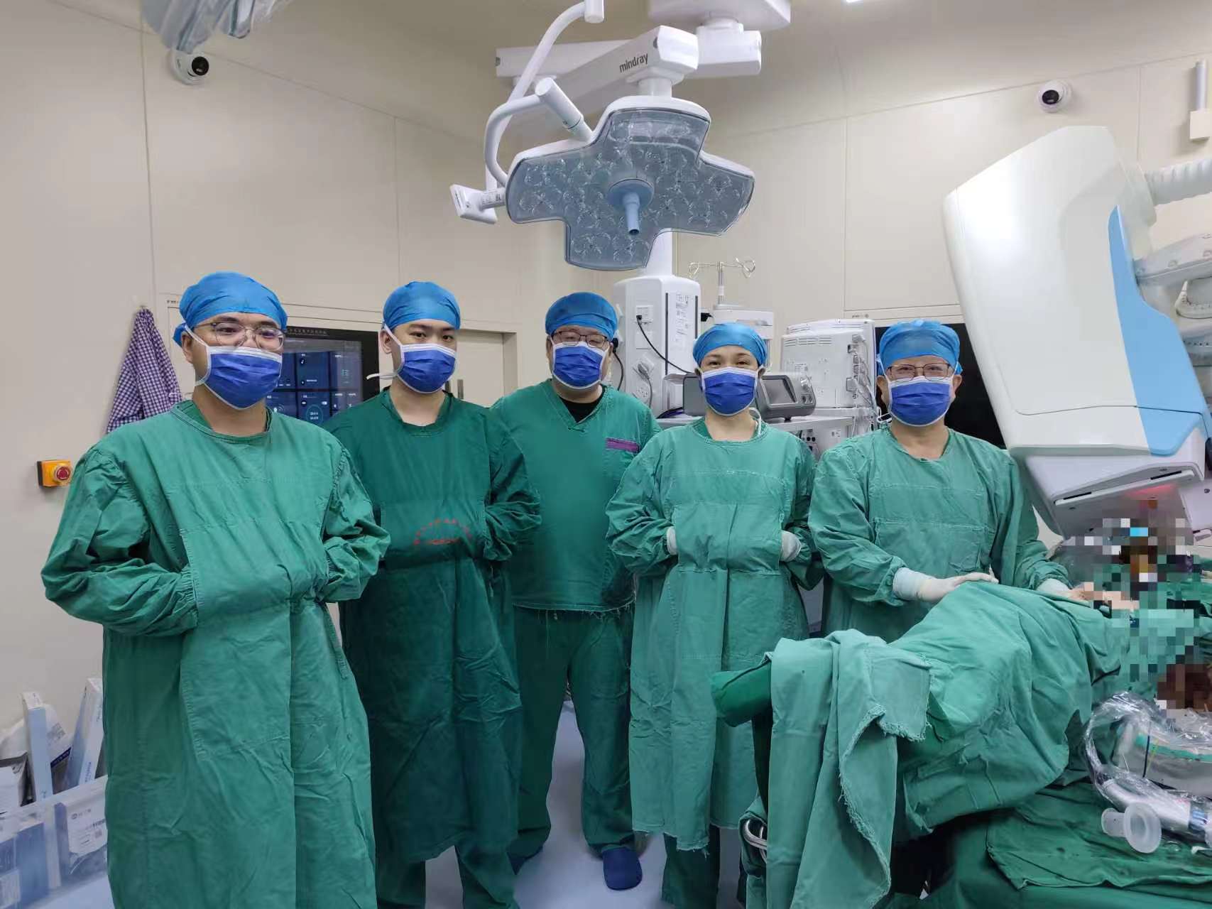
On August 10, 2026, The First Affiliated Hospital of University of South China used HC-IORT to successfully complete the world’s first laparoscopic IORT procedure for rectal cancer. Three treatment fields were delivered: two laparoscopic fields and one spherical field. Laparoscopic treatment field 1 — prescribed dose: 12 Gy; energy: 6 MeV; field diameter: 30 mm; field angle: 30°; beam-on time: 3.93 minutes. Laparoscopic treatment field 2 — prescribed dose: 12 Gy; energy: 6 MeV; field diameter: 30 mm; field angle: 45°; beam-on time: 4.28 minutes. Spherical treatment field — prescribed dose: 12 Gy; energy: 9 MeV; field diameter: 40 mm; beam-on time: 4.51 minutes.
This page is provided solely to present clinical application updates and does not constitute medical advice, a basis for diagnosis or treatment, or a commitment regarding product use.